Yao Zhu, Wanjiang Zhang, Siguo Liu, Stefan Schwarz
J Antimicrob Chemother.2021 Jan 11;dkaa555.doi: 10.1093/jac/dkaa555.Online ahead of print.

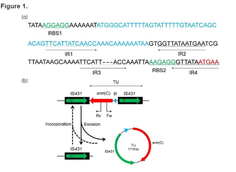
(a) Presentation of the erm(C) translational attenuator. The putative ribosome-binding sites (RBS1 and RBS2) are shown in green, the reading frame for the small regulatory peptide is shown in blue and the start of the erm(C) gene is shown in red. Moreover, the four IRs (IR1–IR4) are indicated by arrows. The deleted 3 bp in IR3 is displayed as hyphens. (b) Schematic presentation of excision and incorporation of the erm(C)-carrying TU from and into the backbone of plasmid pSA01-tet. The IS431 elements are displayed as black boxes with the green arrow inside representing the transposase gene. The erm(C) gene is shown as a red arrow and the small reading frame for the regulatory peptide within the translational attenuator is indicated by a blue arrow. The arrowheads indicate the direction of transcription of the respective genes. The detected TU is shown as a circle. Fw and Rv indicate the positions of the primers used in PCR screening for TUs. This figure appears in colour in the online version of JAC and in black and white in the print version of JAC.
https://academic.oup.com/jac/advance-article/doi/10.1093/jac/dkaa555/6082781